Heterogeneity
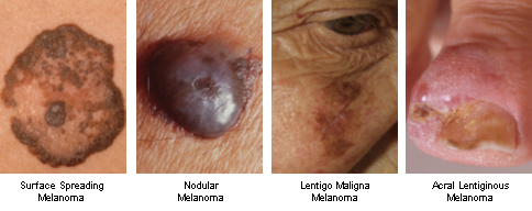
Melanoma is a heterogeneous disease at many different levels. At the clinical level there are four major types of primary lesion and the disease progresses through several well-defined stages. Under the microscope melanoma cells present a variety of morphologies and immunohistochemical analysis frequently reveals non-uniform distribution of various marker proteins within lesions. In the laboratory, different cell lines have distinctly different phenotypes of proliferation, motility, invasion and response to growth factors. Finally, at the molecular level there are a range of mutations, patterns of genomic aberration and at least two well-defined transcriptional states. Thus the different ways of considering the disease have each revealed multiplicity of presentation at all levels.
 |
Heterogeneity in the Hoek lab
The core of our research has become the study of cellular heterogeneity in melanoma. I came to this in 2002/2003 while examining the results of expression profiling (DNA microarray) experiments carried out on in vitro cultures of metastases of human melanoma. I had originally tried to link gene expression with characteristics of patient response to treatment, but strict adherence to the rules of statistical analysis did not reveal any such connections. However, I did notice that there seemed to be sub-groups of gene expression patterns. Frustratingly, these sub-groups did not appear to be associated with any patient characteristic, or with the mutation status of BRAF and NRAS. For a while, I suspected that these sub-groups may simply be an artifact typical of class-discovery analyses of high-throughput data. It was an uncomfortable feeling to think I was chasing a red herring.
I then took the opportunity to perform the same class-discovery analyses on two additional datasets, supplied by the laboratories of Meenhard Herlyn and Dirk Schadendorf. As expected, I also found sub-groups. The critical characteristic of this is that the genes involved, whose expression variations underlie those sample sub-groupings, were the same in all cases. I had found what Colin Goding would later describe as a taxonomy of melanoma cells. Unfortunately, this taxonomy did not seem to be connected with anything. From Meenhard's data we could see that sub-group membership had nothing to do with whether cells came from primary or metastatic samples. From Dirk's data we could confirm that sub-group membership was not connected with any patient characteristic, nor with a range of gene mutations. So while we had convinced ourselves the sub-grouping could not be an artifact we didn't know what it meant.
In the lab I started to explore what this taxonomy was all about. It was clear from the microarray data that one sub-group expressed all the melanocytic markers that were expected for melanoma (Mitf, Tyr, melan-A, etc), and that the other sub-group did not. An early finding was that melanocytic sub-group cultures were pretty easy to grow, but the non-melanocytic samples were slower cycling (sometimes very slow cycling) and took longer to grow enough for in vitro studies. A second connection between sub-grouping and biology was made with the help of Natalie Schlegel, our first PhD student. We found that the sub-group which expressed melanocytic markers was significantly more susceptible to growth inhibition by TGF-beta.
Traditionally, susceptibility to growth-inhibition by TGF-beta has been held to be a characteristic of early phase cancer cells (including melanoma). It was thought that part of cancer progression involves overcoming this susceptibility, that susceptible early phase cells had weak metastatic potential and that resistant late phase cells had strong metastatic potential. For us this did not make any sense at first, because our sub-groups (we had started to call them 'cohorts' by then) had nothing to do with whether or not a sample was derived from an early phase (e.g. primary) or a late phase (e.g. metastatic) melanoma. This is when I had what I think of as my One Good Idea (everybody get's one, I suppose), and this was it:
| We note that immunohistochemical analyses of melanoma biopsies, using antibodies targeting the products of genes which we mention, often yield heterogeneous staining patterns among otherwise morphologically similar cells. This suggests to us that individual melanoma growths are comprised of cells from across the cohort spectrum, including populations which are proliferative but less metastatic than others less proliferative and yet more invasive, TGF-beta-resistant, and secreting factors which change microenvironmental architecture and encourage neovasculogenesis. We suspect that microenvironmental cues, such as hypoxia and inflammation, may allow melanoma cells to switch epigenetically between cohort transcriptional signatures. |
In other words, melanoma progresses as a disease because melanoma cells can switch back-and-forth between states of proliferation and invasion and that this switching is regulated by microenvironmental changes.
After quite a long time and many rejection slips from many journals, Colin Goding (then editor-in-chief for Pigment Cell & Melanoma Research) let us publish exactly this in 2006. We've based all of our studies on that idea ever since - although we now refer to it as the phenotype switching hypothesis.
Relevant papers
Hoek et al., 2006
Hoek et al., 2008.
Schlegel et al., 2009
Eichhoff et al., 2010
Hoek & Goding, 2010
Zipser et al., 2011
Eichhoff et al., 2011
Widmer et al., 2012